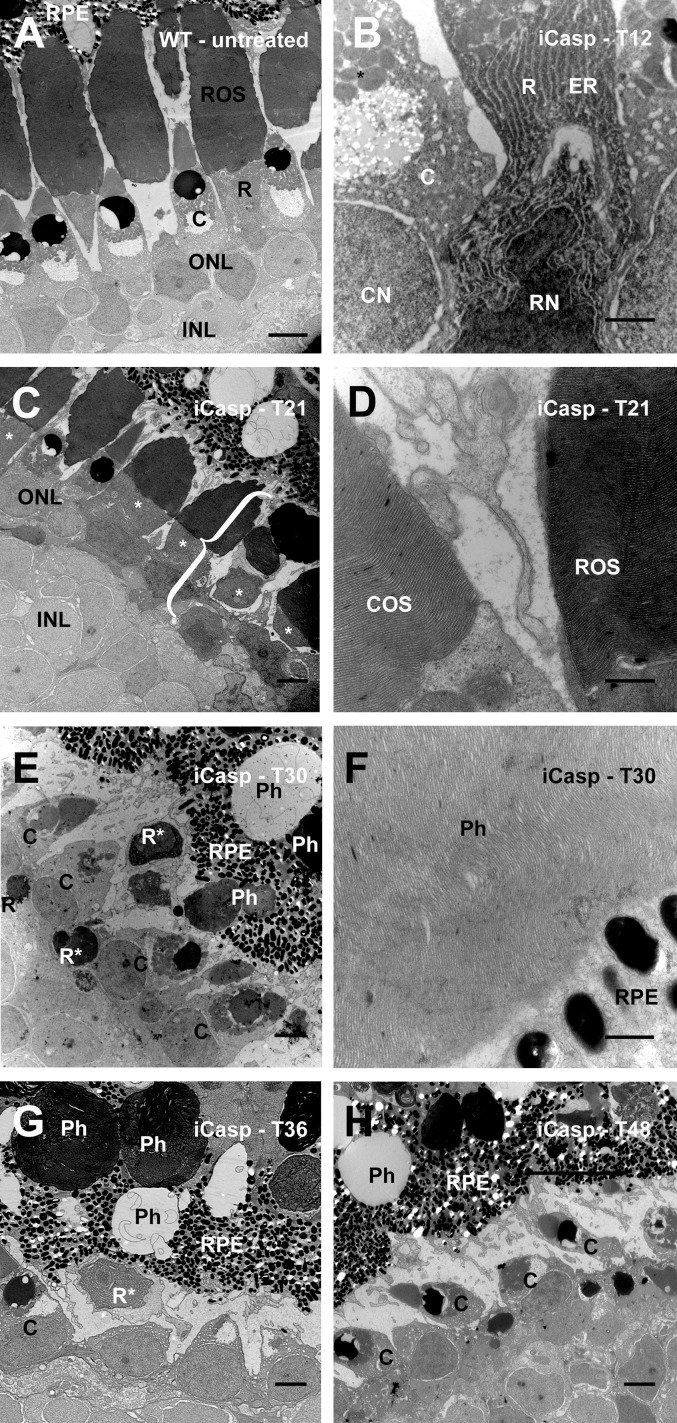
Figure 7.
Rod outer segment vesiculations and increased number of autophagic compartments were not observed in a drug-induced rod apoptosis model. Images are derived from (A) untreated WT control; and (B) iCasp9-expressing tadpole 12 hours after AP20187 administration. There is minimal evidence of cell death, with darkly staining rod cytoplasm and some evidence of altered nuclear morphology. (C) iCasp9-expressing tadpole 21 hours after AP20187 administration. Rods* stained darkly relative to other cell types, and cell fragmentation was initiated (bracket). (D) Rod and cone outer segment (OS) disks retained a high degree of organization at 21 hours. (E) iCasp9-expressing tadpole 30 hours after AP20187. Numerous apoptotic rod remnants are present, and many are undergoing phagocytosis by the RPE. (F) Rod OS remnants in RPE phagosomes retain a high degree of disk organization. (G) iCasp9-expressing retina 36 hours after AP20187 administration. Rod remnants are present in phagosomes of the RPE. (H) iCasp9-expressing retina 48 hours after AP20187 administration (essentially rodless). C, cone; Ph, phagosome; R, rod; R*, apoptotic rod fragment. Scale bars: 10 μm (A, C, E–H), 2 μm (B), 500 nm (D, F).

