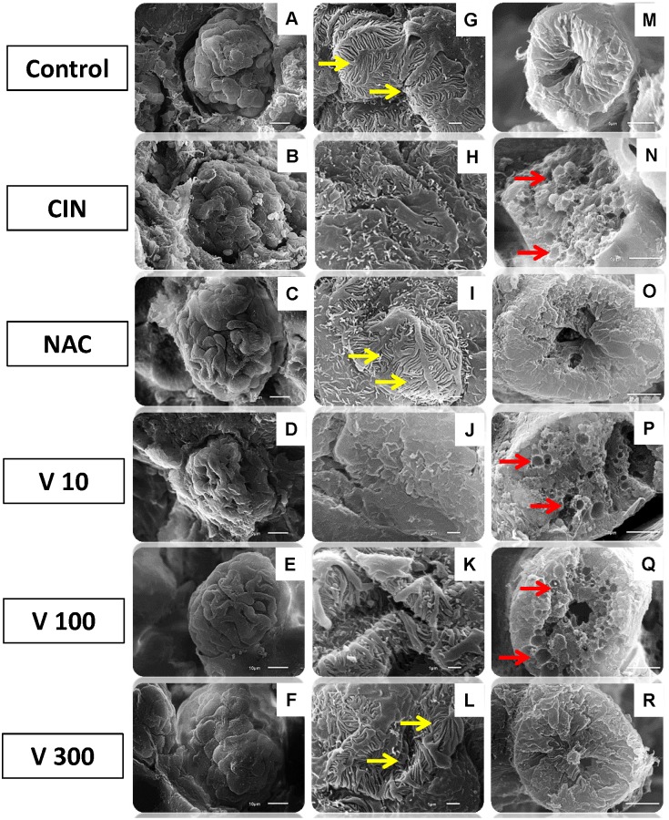
Fig 4. Scanning electron microscopy (SEM) photographs.
Freeze-fractured kidney tissue samples from different groups confirming the decrease in renal glomerular and tubular injuries. The first column (A-F, scale bar = 10 μm) shows scanning images of whole glomeruli, showing structural preservation of the surface tissues in the NAC (C) and V 300 (F) groups, similar to the Control group. The CIN (B), V10 (D) and V100 (E) groups exhibited shrunken glomerular tufts accompanied by loss of structural cohesion. The second column (G-L, scale bar = 1 μm) shows higher magnification of podocytes to display the primary processes and the interdigitating secondary processes. The CIN (B), V10 (D) and V100 (E) groups showed atypical podocytes. Only the NAC (I) and V300 (L) groups were similar to the control structures (G), with smooth foot processes that tightly apposed each other (yellow arrows). The third column (M-R, scale bar = 5 μm) shows scanning images of proximal tubules with normal structure (M), with vacuolization and luminal congestion (red arrows) after radiocontrast nephropathy (N, P and Q) and attenuation of cytoplasmic vacuoles in the NAC (O) and V300 (R) groups.

