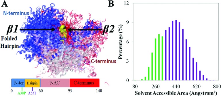
FIG. 5.
β-hairpin interacting with α-synuclein C-terminus. (a) Overlay of structures of simulated α-synuclein containing β-hairpin formed in region 38-53. The structures are aligned at the β-hairpin region shown in gold (region 38-53). N-terminus, β-hairpin region, NAC region, and C-terminus are colored blue, gold, pink, and red, respectively. Shown beneath the structure is a schematic depiction of the α-synuclein segments as defined in Fig. 1. (b) Distribution of solvent-accessible surface area (SASA) of β-hairpins in region 38-53. The distribution is colored green and purple to distinguish buried and exposed β-hairpin conformations, respectively.

