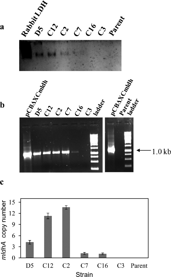
Fig 2. Analysis of mldhA transformants by activity staining, genomic PCR and qPCR: a—Activity staining for LDH from A. niger and six of its transformants.

Desalted cell-free extract (5 μg protein each) was loaded on polyacrylamide gels. Purified rabbit skeletal muscle LDH served as positive control. b—PCR amplification of integrated PcitA- mldhA DNA from the transformants. Parent A. niger genomic DNA and pCBXCmldh served as negative and positive controls, respectively.c—PcitA-mldhA copy number determination in transformants by qPCR. The mldhA copy number was determined using the single copy A. niger actin gene as reference. The data are plotted as mean values obtained from different concentrations of genomic DNA. Error bars represent the standard deviation.
