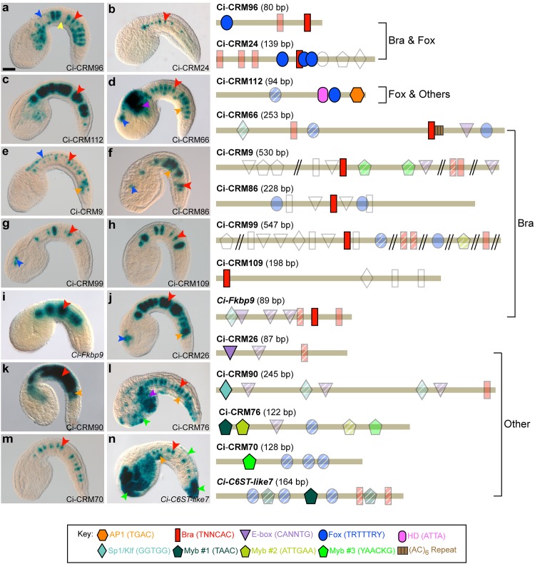
Fig 1. A comparative study of notochord CRMs in Ciona.
a-n: Microphotographs of transgenic Ciona embryos expressing the LacZ reporter in the notochord (red arrowheads) under the control of 14 CRMs. (Right) Schematic representations of the 14 minimal notochord CRMs. Putative transcription factor binding sites are mapped along the length of each enhancer (tan bar), as indicated in the key (bottom). Point mutations uncovered site(s) required for notochord expression (colored and opaque) as well as sites that did not evidently contribute (colored, but transparent). Putative binding sites deemed dispensable through truncations are colored and hatched. Untested putative sites are outlined in gray. Additional staining domains are indicated by arrowheads, colored as follows: blue: CNS, yellow: endoderm, orange: muscle, purple: mesenchyme, green: epidermis. Embryos are oriented with dorsal up and anterior to the left. Scale bar: 40 μm. See also S1, S2 and S3 Figs

