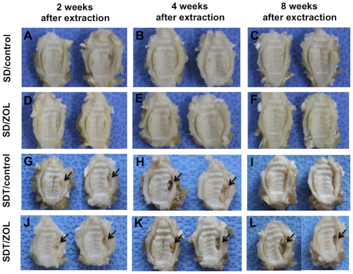
Fig 3. Macroscopic view of extraction sockets in SD/control (A-C), SD/ZOL (D-F), SDT/control (G-I), and SDT/ZOL (J-L) rats.

Two representative specimens are shown. Normal healing after molar extraction was observed in SD/control (A) and SD/ZOL (D) rats by 2 weeks after extraction. Extraction sockets in both groups were covered with intact epithelium at 4 weeks (B, E) after extraction and at 8 weeks (C, F) after extraction. Apparent mucosal disruption with exposed bone (arrows) was seen at the extraction site at 2 (G) and 4 (H) weeks, but not at 8 weeks (I) after extraction in the SDT/control group and at 2 (J), 4 (K), and at 8 weeks (L) after extraction in the SDT/ZOL group.
