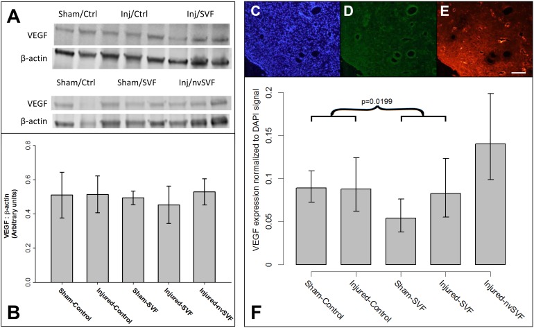
Fig 4. (A,B) Expression of vascular endothelial growth factor (VEGF) and β-actin measured by Western blot revealed no significant differences across experimental groups. Representative images of endometrial DAPI (C), green fluorescent protein (D), and rhodamine (E) expression measured by immunohistochemistry in an injured-control horn. A significant reduction in VEGF expression normalized to DAPI was collectively noted across experimental groups that received SVF treatment (asterisk), though no significant differences were noted between individual groups (F).
Error bars represent standard deviation. Scale bar = 100 μm.

