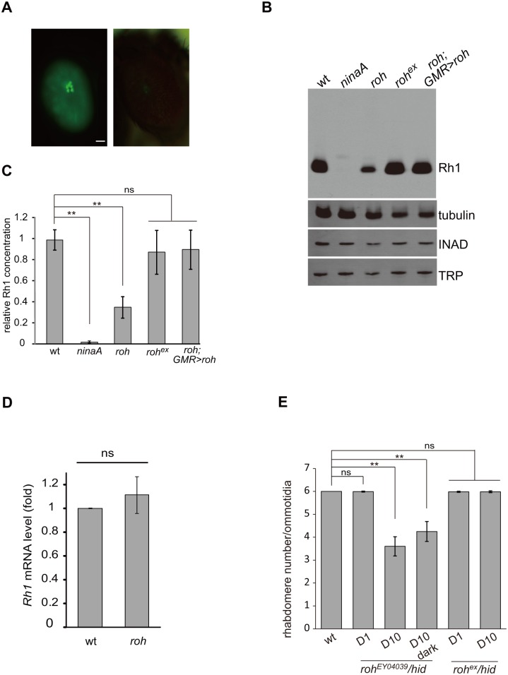
Fig 6. roh mutation results in Rh1 decrease.
(A) GFP fluorescence of one-day old wild-type (left panel) and roh EY04039 (right panel) flies showing a reduction in Rh1-GFP. Scale bar, 50 μm. (B) Western blotting showing decreased Rh1 levels in the roh EY04039 mutant (ey-flp Rh1::GFP; FRT42D roh EY04039 / FRT42D GMR-hid CL); the INAD and TRP levels are not affected compared to the wild-type. The reduction of Rh1 levels could be rescued in roh ex (ey-flp Rh1::GFP; FRT42D roh ex /FRT42D GMR-hid CL) and roh EY04039;GMR>roh (ey-flp Rh1::GFP; FRT42D roh EY04039 / FRT42D GMR-hid CL;GMR-gal4/UAS-roh) flies. Flies less than 1 day old were used. (C) Quantification of relative Rh1 level in various genotypes: wt, ninaA 1, roh EY04039, roh ex and roh EY04039;GMR>roh. The Rh1 levels were normalized to tubulin. (D) Quantitative real-time PCR of wild-type (ey-flp Rh1::GFP; FRT42D/ FRT42D GMR-hid CL) and roh EY04039 fly head with rh1-specific primers. The results are normalized to the expression level of gpdh. The graphs represent the means ± SD of three independent experiments. Error bars represent the SD. (E) Average rhabdomere numbers per ommatidia of the roh EY04039 mutant under the indicated conditions. Each data point was based on examination of >60 ommatidia from >3 flies. Error bars represent the SDs. Asterisks indicate statistically-significant differences (one-way ANOVA and post-hoc Dunnett’s test; ns: not significant, **p < 0.01).

