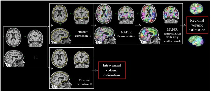
Fig 1. Pincram brain extraction and MAPER segmentation.
MRI T1 sequences were processed with pincram, followed by MAPER and grey matter masking to obtain regional grey matter volumes. Extraction P was used to estimate the intracranial volume. Pincram extraction H: Brain extraction using the Hammers atlases; Pincram extraction P: Brain extraction using the PACO-customized atlases.

