Figure 1.
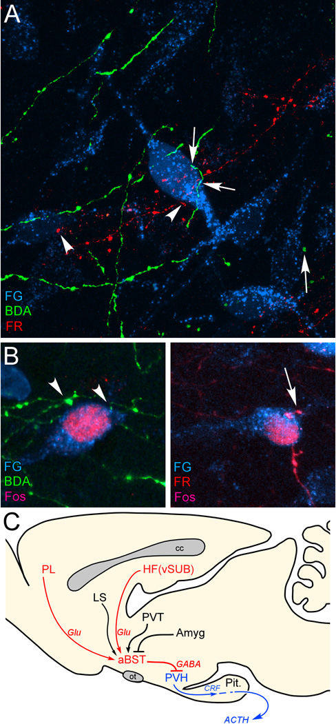
A: Confluence of labeling in aBST following retrograde tracer injections in PVH (Fluoro-Gold, FG; cyan), and anterograde tracers in PL (biotinylated dextran amine, BDA; green) and vSUB (FluoroRuby, FR; red). Instances of BDA- (arrows) and FR-labeled (arrowhead) terminals make appositions onto single PVH-projecting neurons in aBST, by laser-scanning confocal microscopic analysis. B: After a single stress exposure, numerous instances of Fos-labeled nuclei are evident in PVH-projecting neurons containing appositions from BDA- (left) and FR-labeled (right) terminals. C: Previous work of ours supports the pathways highlighted in red, with aBST providing an important source of GABAergic innervation of PVH, and relaying limbic cortical influences. Other forebrain cell groups known to influence HPA output (highlighted in black) also project to aBST, whose integrated output targets PVH directly. Like vSUB and PL, these regions do not provide any appreciable innervation of PVH, but do issue projections to the aBST. ACTH, adrenocorticotropic hormone; Amyg, amygdala; cc, corpus callosum; CRF, corticotropin-releasing factor; Glu, glutamate; HF, hippocampal formation; LS, lateral septum; ot, optic tract; Pit., pituitary gland; PL, prelimbic cortex; PVH, paraventricular nucleus of the hypothalamus; PVT, paraventricular thalamic nucleus; vSUB, ventral subiculum. Data are based upon Radley and Sawchenko (2011).

