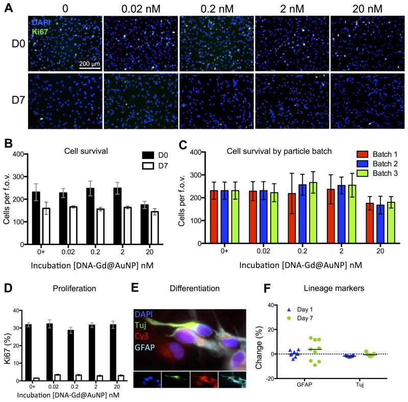
Figure 4. Cellular effects of DNA-Gd@AuNPs.
Cells at day 0 and day 7 after labeling were stained with DAPI and Ki67 (A, scale bar = 200 μm). No significant effects were seen on the number of surviving cells at any concentration of DNA-Gd@AuNPs at day 0 or day 7 (B) and this effect remained consistent when three separate batched of particles were tested (C). The percentage of cells expressing Ki67 also remained consistent at all DNA-Gd@AuNP concentrations at day 0 and day 7 (D). Cells were also stained to assess phenotypic changes (E), and no significant differences were seen between those labeled at 20 nM nanoparticles and controls (F).

