Fig. 11.
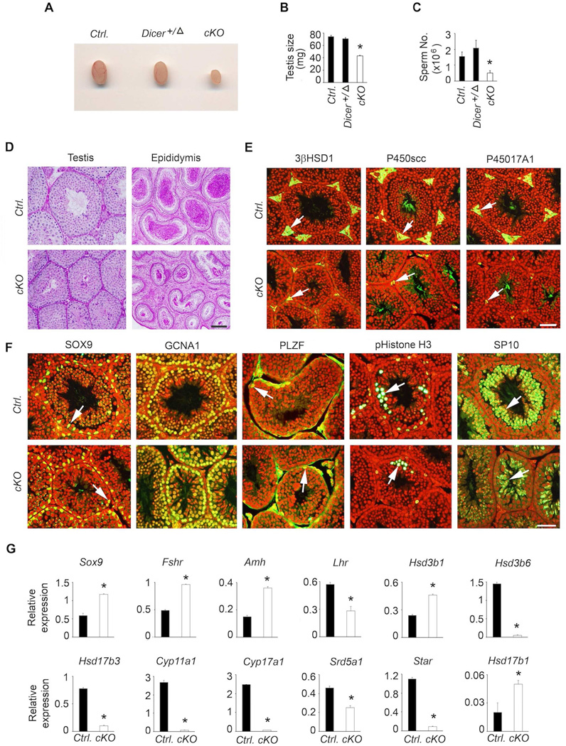
Analysis of male reproductive phenotypes in Dicer cKO mice indicate that grossly (A) testis size (B) and sperm number (C) are decreased compared to those in age-matched littermate controls. Histological analysis of testis by PAS/hematoxylin staining shows decreased testis tubule size and reduced sperm number in cauda epididymis (D), immunolocalization reveals hypoplasia of Leydig cells (white arrows in E) and grossly normal expression of Sertoli and germ cell markers (F), but real time qPCR assays indicate aberrant testis somatic cell gene expression (G). For each experiment, at least 4–5 adult mice at 42 days of age are used. Real time qPCR assays are done in triplicate per sample. In A–C, * P < 0.01 compared to corresponding controls by One way ANOVA followed by Turkey’s post-hoc test. For qPCR assays, * P < 0.01 by T-test. In E and F panels, merged images are shown and nuclei are stained red. Bars in D-F = 100 µm.

