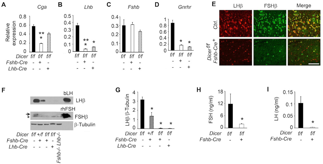
Fig. 12.
Defects in gonadotropin homeostasis in Dicer cKO female mice are analyzed exactly as described in Fig. 10. Real time qPCR assays (A–D), dual immunofluorescence localization (E–G) and serum RIAs (H and I) all confirm that gonadotropin synthesis and secretion are suppressed in Dicer cKO mice compared to age-matched littermate controls. An asterisk below the arrow in (F) indicates a non-specific band. In A-D, and G, * P < 0.01 compared to corresponding controls and in A, ** P < 0.01 Dicer deletion with Fshb-Cre vs. Lhb-Cre by One way ANOVA followed by Turkey’s post-hoc test. In H and I, P < 0.01 by T-Test. Adult female mice at 6 weeks of age (n=4–5) are used in each experiment. Representative immunolocalization (E) and immunoblots (F) are shown. White bar in E = 100 µm.

