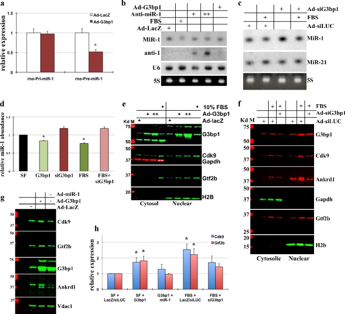
Fig 3. G3bp1 regulates miR-1 processing in cardiomyocytes.
(a) Neonatal myocytes were treated with Ad-LacZ or Ad-G3bp1 for 24hrs; total RNA extracted was used for qPCR analysis for rno-primary and rno-pre-miR-1. The results were normalized to 18S, averaged and plotted. Error bars represents SEM, and * is p<0.05, n = 3. (b) Neonatal myocytes cultured in serum free conditions were treated with Ad-LacZ, Ad-G3bp1, Ad-anti-miR-1 (two doses, as indicated) or 10% FBS for 24hrs before extracting total RNA for Northern Blots. Blots were probed for miR-1, anti-miR-1 sequence or U6. 5S is shown as loading control. (c) Myocytes were cultured and treated with adenovirus expressing shRNA against G3bp1 (Ad-siG3bp), Ad-siLUC (control virus) or 10% FBS, as indicated. Northern blots were performed and probed for miR-1 and miR-21 (specificity control), 5S is shown as loading control. (d) Northern blots from 3a and b, and two independent blots were scanned, quantified, averaged and plotted. The error bars represents SEM and * p<0.05 vs. SF control. (e) Western blot analysis of the cytoplasmic and nuclear fractions of protein lysate was performed on myocytes for the indicated genes on myocytes treated with Ad-lacZ (control virus), Ad-G3bp1 or 10% FBS, as indicated for 24hrs. (f) Western blot analysis of cytoplasmic and nuclear fractions of protein lysate was performed on myocytes treated with Ad-siLUC (control virus), Ad-siG3BP1 or 10% FBS, as indicated. (g) Total protein lysate from cultured myocytes treated with Ad-lacZ (control virus), Ad-G3bp1 or Ad-miR-1, as indicated were separated by SDS-PAGE and protein expression of selected genes analyzed, n = 2. (h) Western blots were quantified, averaged and plotted. Error bars represents SEM and * p<0.05 vs. respective control. All experiments were performed in triplicates, unless indicated otherwise and representative blots have been shown.

