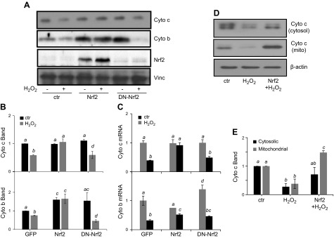
Figure 6.
Nrf2 overexpression attenuates decreases in expression of mitochondrial proteins. Rat cardiomyocytes were infected with adenovirus for expression of GFP, Nrf2, or DN-Nrf2. At 48 h after infection, cells were treated with 300 μM H2O2 for 2 h and were collected at 48 h after H2O2 treatment for measurements of Nrf2, cytochrome c, and cytochrome b by Western blot (A, B) or real-time RT-PCR (C). Cytosolic and mitochondrial fractions were purified as described in Materials and Methods for Western blot analysis of cytochrome c protein levels (D, E). The band intensity from Western blot was quantified using ImageJ software and normalized to the corresponding loading control VCL or β-actin band for numeric presentation (B, E). Values represent the means ± sd from 3 independent experiments. A letter a indicates significant difference (P < 0.05) from means labeled with a letter b, c, or bc as determined by 1-way ANOVA with Bonferroni correction. The mean labeled ac or bc is not significantly different from that labeled with a or c, or b or c, respectively.

