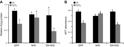
Figure 8.
Nrf2 prevents the decrease of mitochondrial function from H2O2 exposure. Neonatal rat cardiomyocytes were infected with adenovirus for expression of GFP, Nrf2, or DN-Nrf2. At 48 h after infection, cells were treated with 300 μM H2O2 for 2 h. At 5 d after H2O2 exposure, cells were stained using MitoTracker Orange CMTMRos for quantification of membrane potential (A). Fluorescence intensity was normalized to protein content from the corresponding well (A). Mitochondrial succinate dehydrogenase activity was assessed by incubation of cells with MTT (B). Values represent the means ± sd from 3 independent experiments. A letter a indicates significant difference (P < 0.05) from means labeled with a letter b as determined by 1-way ANOVA with Bonferroni correction.

