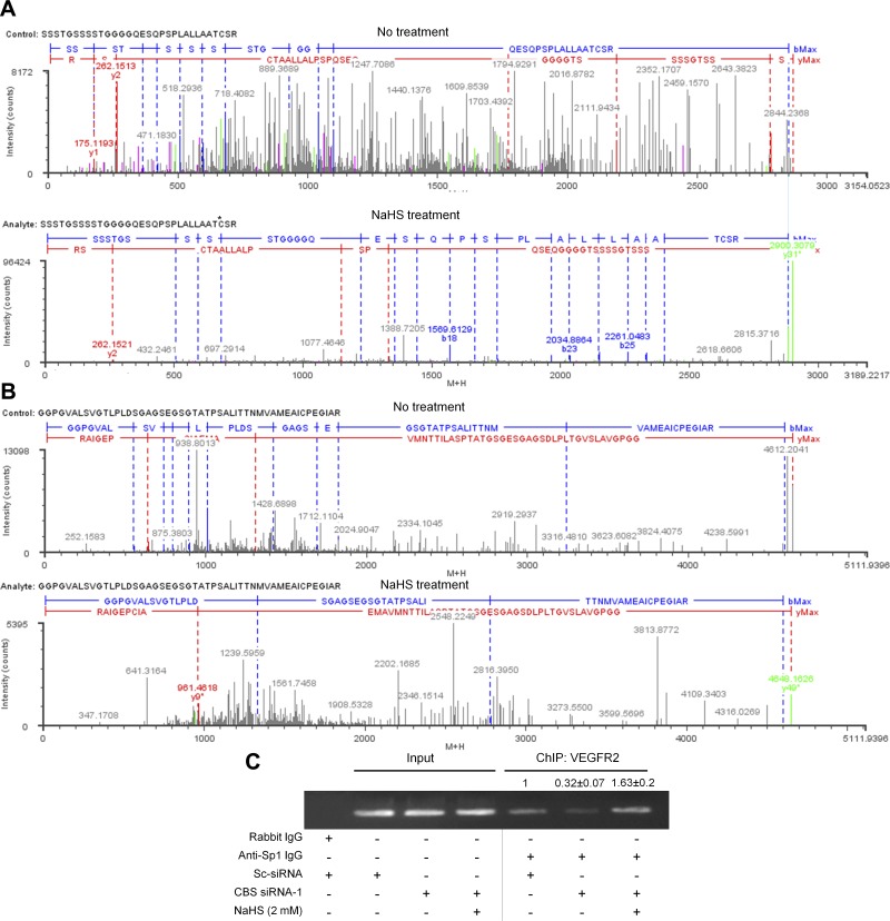
Figure 8.
A, B) Charge-reduced, isotope-deconvoluted MSE spectra (fragment ion matched profiles) of untreated and 100 µM NaHS-treated full-length Sp1 protein. *The site of sulfhydration. C) ChIP assay to determine relative Sp1 binding to the VEGFR-2 promoter in scrambled or CBS siRNA- or CBS siRNA + NaHS (2 mM)-treated HUVECs.

