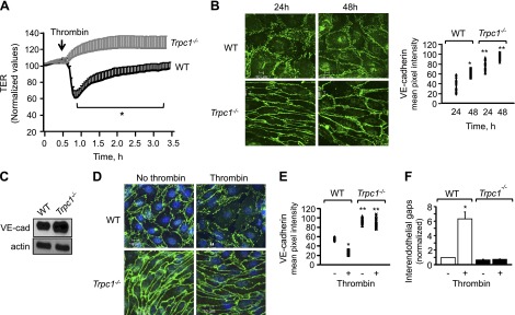
Figure 1.
Deletion of TRPC1 augments cell-surface VE-cadherin expression, which stabilizes AJs and prevents increase in endothelial permeability by thrombin. A) Changes in TER after stimulation of indicated ECs with thrombin. *Values lower than Trpc1−/− ECs; P < 0.05. B) In parallel, ECs were stained with anti–VE-cadherin antibody to determine AJs integrity. Top, Representative image of AJs at indicated times. Bottom, Mean ± sd of VE-cadherin cell surface pixel intensity; n = 3 per group. *Values higher than WT ECs at 24 h; **values higher than WT ECs at each of indicated times; P < 0.05. C) VE-cadherin protein expression in indicated ECs using anti-VE-cadherin antibody. Actin was used as loading control. Immunoblot represent data from experiments repeated at least 3 times. D–F) VE-cadherin cell-surface intensity and interendothelial gap formation after 5 min thrombin challenge of WT or TRPC1-null ECs. D) Representative image. E, F) Means ± sd of VE-cadherin pixel intensity and interendothelial gap formation from at least 10 individual cells per experiment. Each experiment was repeated at least 3 to 4 times. *Values different than WT ECs without thrombin or Trpc1−/− ECs; **values different than WT EC after thrombin stimulation or no thrombin stimulation; P < 0.05.

