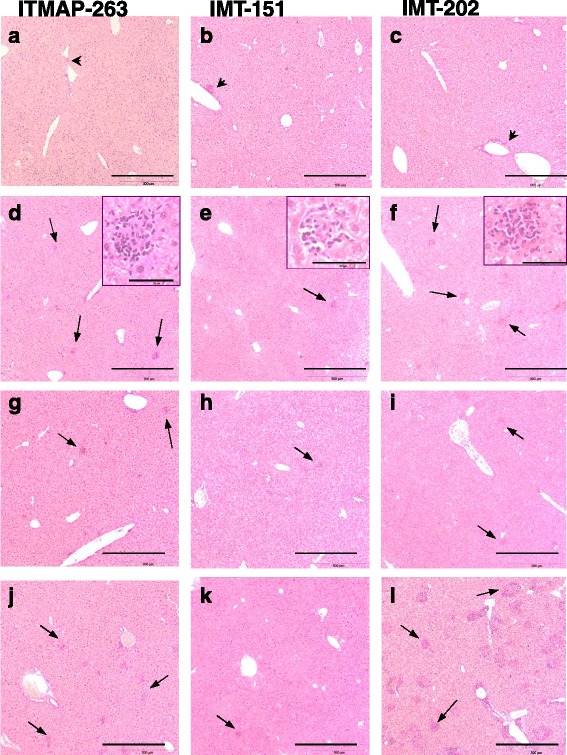
Fig. 4.

Histological alterations in the livers of mice infected with L. infantum. C57BL/6 (a-f), p47phox −/−(g-i) or Nos2 −/−(j-l) mice were infected with L. infantum ITMAP-263 (a, d, g, j), IMT-151 (b, e, h, k) or IMT-202 (c, f, i, l). Liver sections were collected 15 (a-c) and 60 (d-l) days after infection, processed and stained with hematoxylin-eosin. Representative images of each experimental group are shown. Note the inflammatory infiltrates close to the vessels at 15 days post-infection (a-c, blackarrow heads) and throughout the liver parenchyma at day 60 post-infection (d-l, black arrows). Insets in d-f depict in a higher magnification one typical granuloma representative of each parasite strain. The bar corresponds to 500 μm, except for the insets in d-f in which the bar corresponds to 50 μm
