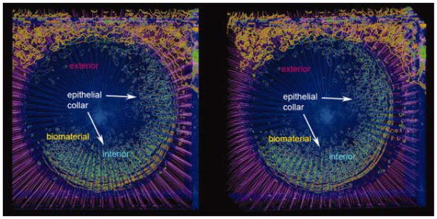
Figure 5.
Stereo pair representing the 3D reconstruction of analyzed micrographs through the region of epithelial incorporation as viewed from the ventral aspect. Green depicts the contours surrounding keratin stain within the porous biomaterial. Keratin stain contours (hair follicles and epidermis) outside the biomaterial are depicted in orange. Red dots depict the intersection between radial lines and keratinocyte stain contours and represent the epithelial migrating front. Radial lines within the biomaterial are represented in blue while radial lines outside the biomaterial are magenta.

