Figure 1.
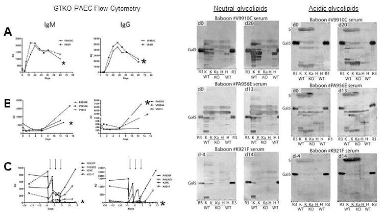
Antibody responses to GalT-KO PAECs and glycolipids after pig-to-baboon GalT-KO:CD55/59/39/H-transferase kidney xenotransplantation. Left: Binding of serum IgM and IgG to GalT-KO donor PAECs (left) A; Control not immune suppressed recipients. B; Immune suppressed recipients. C; Immune suppressed recipient further treated with plasmapheresis (arrows). Right: The corresponding antibody (IgG + IgM) reactivity for each of these recipients to neutral and acidic glycolipids from GalT+) and GalT-KO (KO) pig individuals separated on thin-layer chromatography plates. Glycolipids from different pig kidneys were applied (lanes K and Ka) and pig hearts (lanes H) together with reference Galα3nLc4 glycolipid (lane R3). Anti-PAEC profiles for the individual animals tested in the TLC immmunostainings are indicated by asterix to the left. Recipients V9910C (A) and PA956E (B) show a strong to moderate increase in nonGal IgM and IgG while recipient K921F (C) shows minimal induction of anti-nonGal antibody. Adapted from Le Bas-Bernardet et al. (25).

