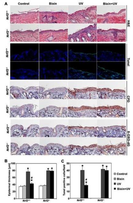
Figure 4. Systemic administration of bixin suppresses UV-induced epidermal thickening, apoptosis, and oxidative DNA damage in Nrf2+/+ mice but not Nrf2−/− mice.
Mice (Nrf2+/+ and Nrf2−/− mice; n = 6 per group) received bixin treatment (200 mg/kg; i.p.) or carrier control (corn oil), followed by solar UV (UVB 240 mJ/cm2) or mock exposure performed 48 h after bixin. (A) After irradiation (24 h), H&E staining and in situ TUNEL analysis visualizing epidermal apoptotic cells were performed [n = 6; representative tissue from each group is shown (scale bar: 100 μm)]. In addition, 8-oxo-dG- and CPD-lesions were visualized by IHC; representative tissue from each group is shown. (B) Epidermal thickness in H&E-stained sections was measured as the distance between the top of the basement membrane and the bottom of the stratum corneum at five randomly selected fields from each mouse specimen. (C) Quantification of TUNEL-positive cells (green fluorescent nuclei) in five random fields per section; 200 × magnification; [means ± SD (*p<0.05, control vs. treatment groups; #p<0.05, UV vs. bixin+UV groups)].

