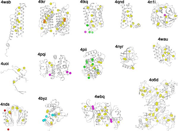
Figure 2. De novo native-SAD structures in 2014.
Structures are identified by associated PDB codes and the contents of the asymmetric unit are shown for each. Protein backbones are represented as ribbon drawings and anomalous scatterers are shown as spheres. Yellow: sulfur; orange: phosphorus; magenta: calcium; green: chloride; cyan: potassium, and red: sodium.

