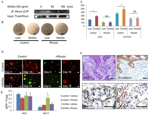
Figure 7. RhoA activation by Wnt5a plays a role in DGC development.
(A) AGS cells were treated with 100 ng/ml Wnt5a for the indicated times. Cell lysates were immunoprecipitated with RhoA-GTP Ab and immunoblotted with total RhoA Ab. (B–C) Soft-agar sphere forming assay of Wnt5a-treated AGS and KATO-III cells. Cells were treated with vehicle or 30 μM Rhosin. Sphere images (B) and numbers (C) of spheres at day 10. n = 4/group. (D–E) Corpus organoids from TAM-treated Mist1-CreERT2; Cdh1flox/flox; R26-mTmG mice treated with 100 ng/ml Wnt5a and/or 30 μM Rhosin. Images (D) and numbers (E) of GFP+Cdh1− organoids per total organoid number on days 3 and 14. n = 4/group. (F) H&E, E-cadherin, Cxcl12, and KLRG1 staining in human DGC. Bars=100 μm (D), 50 μm (B, F). Means ± SEM. *p < 0.05.

