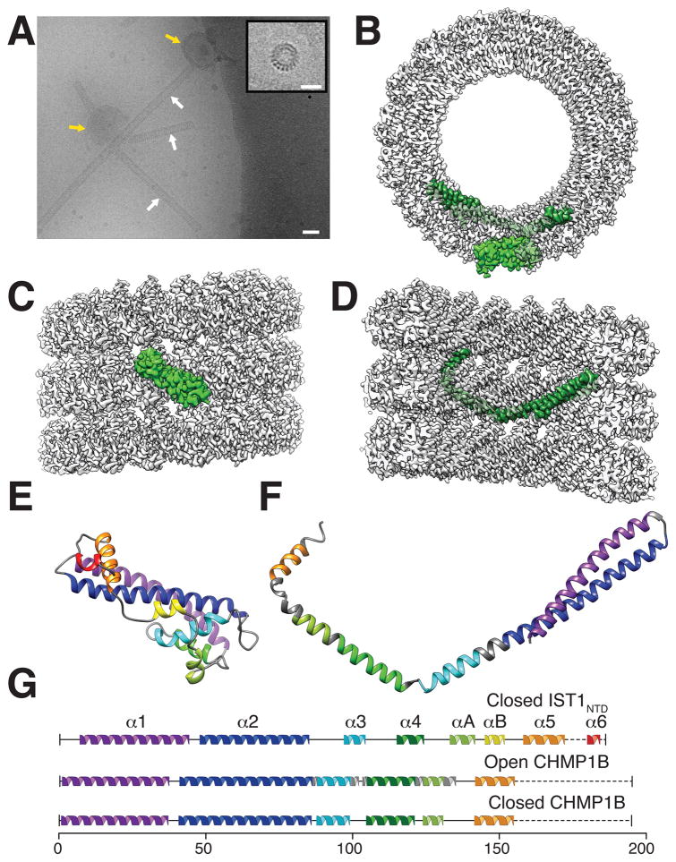
Fig. 1. IST1NTD and CHMP1B copolymerized into helical tubes comprising polar, double-stranded helical filaments.
(A) Electron cryomicrograph showing IST1NTD-CHMP1B tubes (white arrows) assembled by incubating equimolar IST1NTD and CHMP1B in the presence of polymer-nucleating small acidic unilamellar vesicles (SUVs, yellow arrows). Inset: end-on view of a short IST1NTD-CHMP1B tube. Bars: 40 nm (A), 20 nm (inset). (B) End-on view of the reconstructed IST1NTD-CHMP1B tube highlighting single subunits of IST1NTD (light green, outer strand) and CHMP1B (dark green, inner strand). (C) External view of the reconstructed helix with a highlighted IST1NTD subunit. (D) Internal cutaway view of the reconstructed helix with a highlighted CHMP1B subunit. (E) Ribbon diagram of the modeled IST1NTD subunit (closed conformation). (F) Ribbon diagram of the modeled CHMP1B subunit (open conformation). (G) Secondary structure diagrams for closed IST1NTD (top), open CHMP1B (middle), and closed CHMP1B (bottom).

