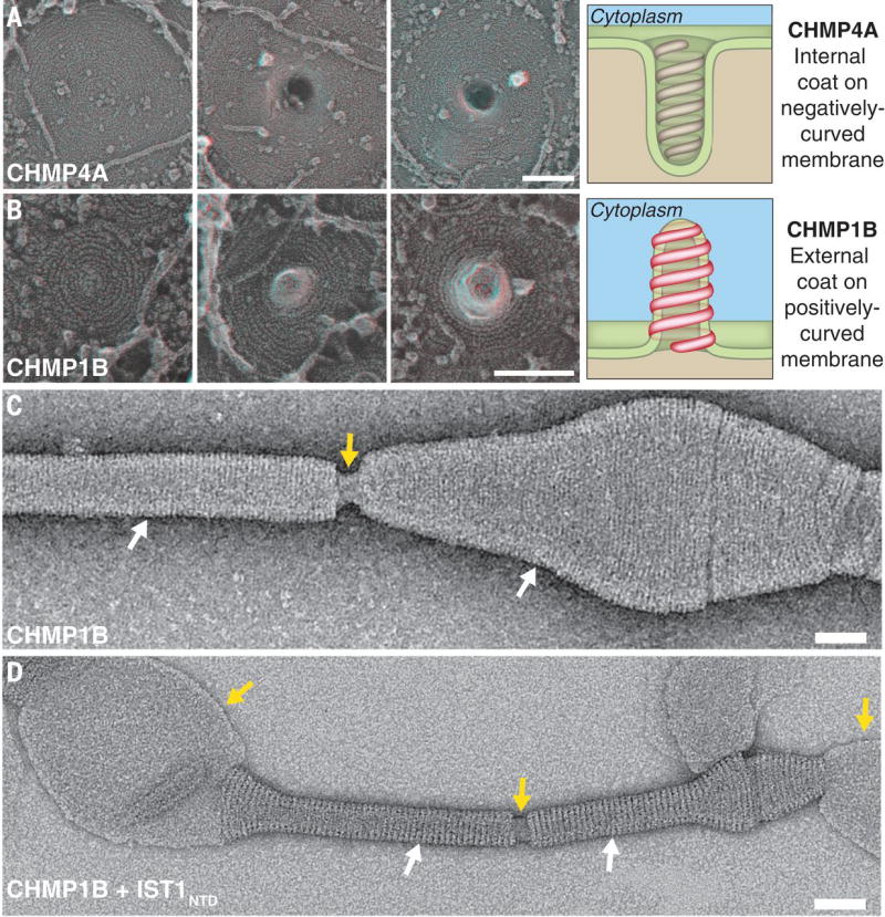
Fig. 4. Topology of ESCRT-III membrane deformation in cells and in vitro.

(A) Series of filament spirals on the plasma membrane of COS-7 cells expressing CHMP4A1–164 show development of the outwardly directed protrusions previously associated with ESCRT-III filaments (15, 16). Drawing highlights relationship between CHMP4A filament spiral and a negatively-curved plasma membrane tubule. (B) Series of filament spirals on the plasma membrane of COS-7 cells expressing FLAG-CHMP1B show development of invaginations directed into the cell. Drawing highlights relationship between CHMP1B filament spiral and a positively-curved plasma membrane tubule. (C) Negative stain electron micrograph showing that CHMP1B tubulates liposomes and forms a filamentous coat on the outside of the tubule. White arrows highlight regions coated by the CHMP1B helices, and the yellow arrow highlights a break in the coat where the internal lipid is visible. (D) Negative stain electron micrograph showing that the IST1NTD-CHMP1B copolymer forms on the outside of membrane tubules. White arrows highlight regions coated by the IST1NTD-CHMP1B copolymer, and the yellow arrows highlight breaks in the helical coat or uncoated regions of the liposome where the internal membrane is visible. Scale bars 100nm (A) and (B), 50 nm (C) and (D).
