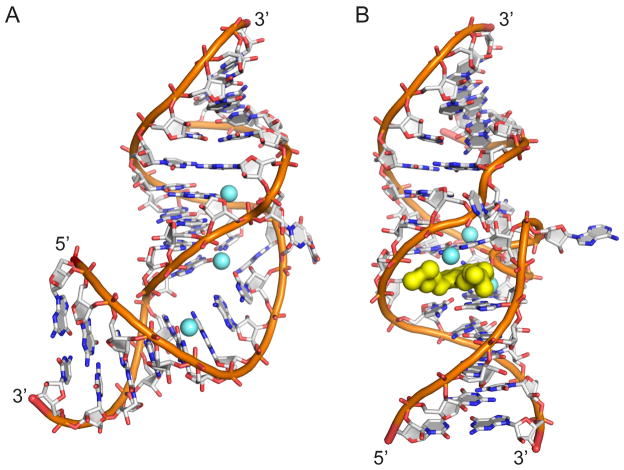
Fig. 1.
Two conformational states of the subdomain IIa RNA switch from the IRES element of HCV as visualized by X-ray crystallography. Both structures contain three magnesium ions (light blue spheres). (A) The RNA switch adopts a bent fold in the ligand-free state. (B) An extended architecture of the RNA switch is captured by benzimidazole viral translation inhibitors and guanine which bind at a deeply encapsulating ligand binding site (yellow surface).

