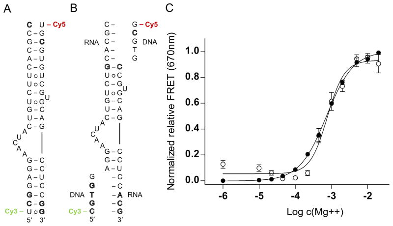
Fig. 2.
Secondary structure and folding of viral RNA switches monitored by FRET experiments with cyanine dye-labeled oligonucleotides. (A) Secondary structure of previously developed, conventional model construct of the HCV IRES subdomain IIa switch 5′ terminally conjugated with Cy3 and Cy5 dyes. (B) Modular FRET construct consisting of unmodified oligonucleotides that contain the HCV RNA switch and carry overhanging single strands which hybridize with cyanine dye-conjugated DNA oligonucleotides. In both panels A and B, outlined letters represent nucleotides deviating from HCV genotype 1b sequence. (C) Normalized relative FRET signal for the Mg2+ titration of the HCV conventional (●) and modular (○) FRET constructs. Dose-response fitting curves gave EC50 values for Mg2+ dependent folding of 730 ± 40 μM (conventional) and 770 ± 80 μM (modular), respectively, for the two different constructs. Error bars represent ±1 s.d. calculated from triplicate experiments.

