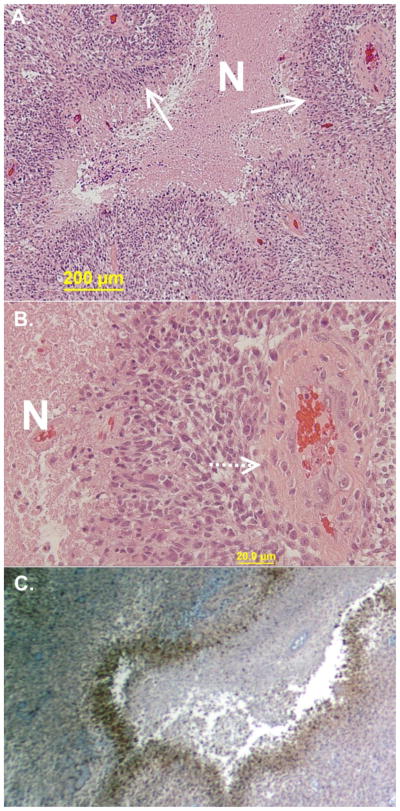
Figure 1.
Representative images of glioblastoma with pathognomonic features and associated hypoxia. A. Highly cellular neoplasm with areas of necrosis (N) with densely packed cells at the margins appearing to almost line up (pseudopalisading). B. Abundant vessels are seen in the tumor with many surrounded by palisades and then necrosis. C. Areas of pseudopalisades are the most hypoxic when stained for the presence of carbonic anhydrase on immunohistochemistry.

