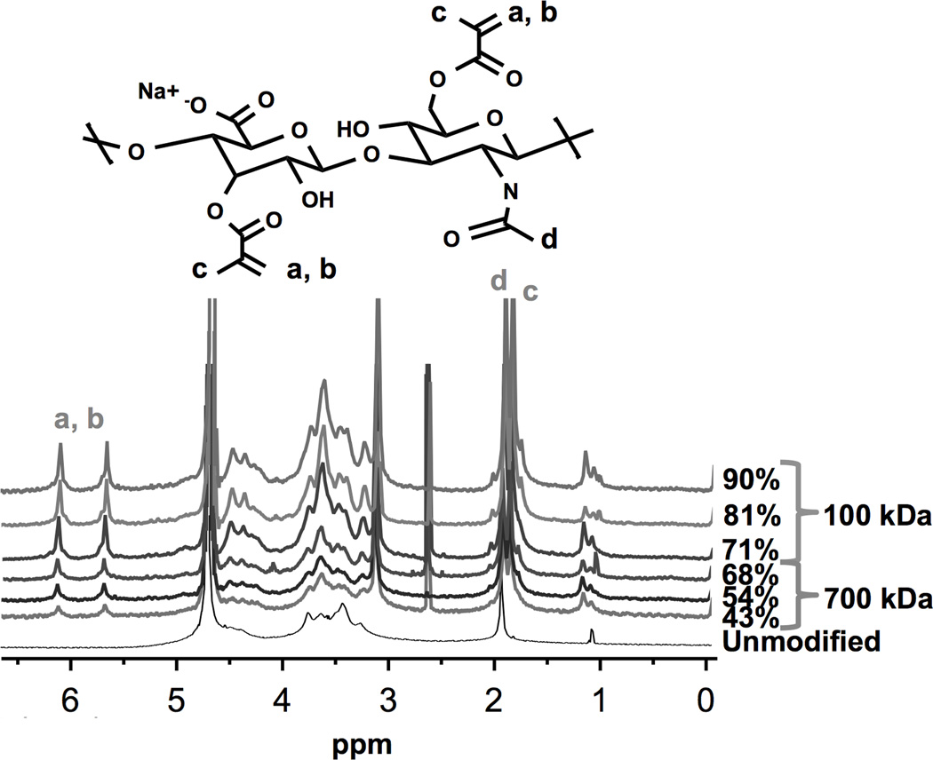
Figure 1.
Chemical structure schematic of methacrylated hyaluronan (HA-MA) repeat unit (top) and 1H-NMR spectra of various HA-MA polymers. Varying degrees of modification (DOM) were achieved through controlled stoichiometric ratios of HA and methacrylic anhydride. The DOM was calculated by taking the ratio of relative integrations of methacrylate peaks (6.1, 5.6, or 1.8 ppm), and HA methyl protons (1.9 ppm). Methacrylate protons at a, b, c and HA’s methyl proton at d are identified in the chemical structure and associated 1H-NMR spectra peaks.

