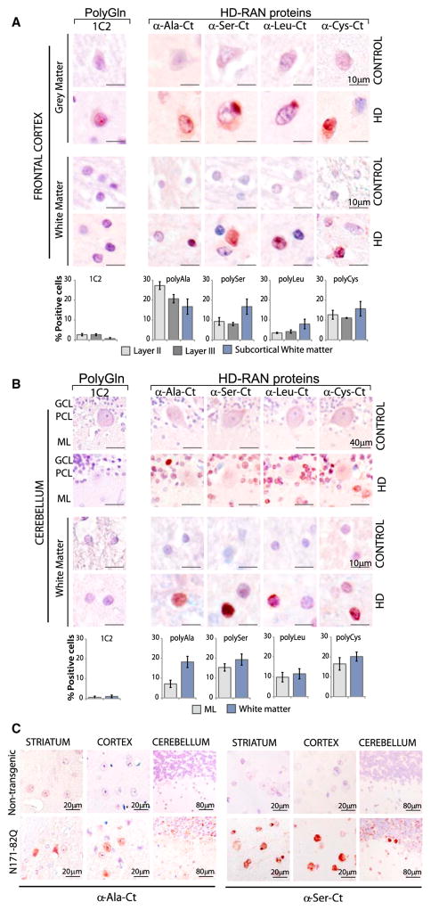
Figure 3. HD-RAN Proteins in Human Frontal Cortex and Cerebellum and HD Mice.
(A and B) IHC staining of control and HD gray and white matter of (A) frontal cortex and (B) cerebellum using α-RAN and α-Gln (1C2) antibodies show punctate nuclear and cytoplasmic staining with α-polyAla, α-polySer, α-polyLeu, and α-polyCys. GCL, granule cell layer; PCL, Purkinje-cell layer; ML, molecular layer. Staining of the cortex and cerebellum in adult-onset HD cases is variable. IHC images and quantification of percent positive cells represent typical positive regions.
(C) IHC staining of indicated brain regions in N171-82Q and control mice using the α-polyAla, α-polySer. Red, positive staining; blue, nuclear counterstain. See also Figures S3A–S3E.

