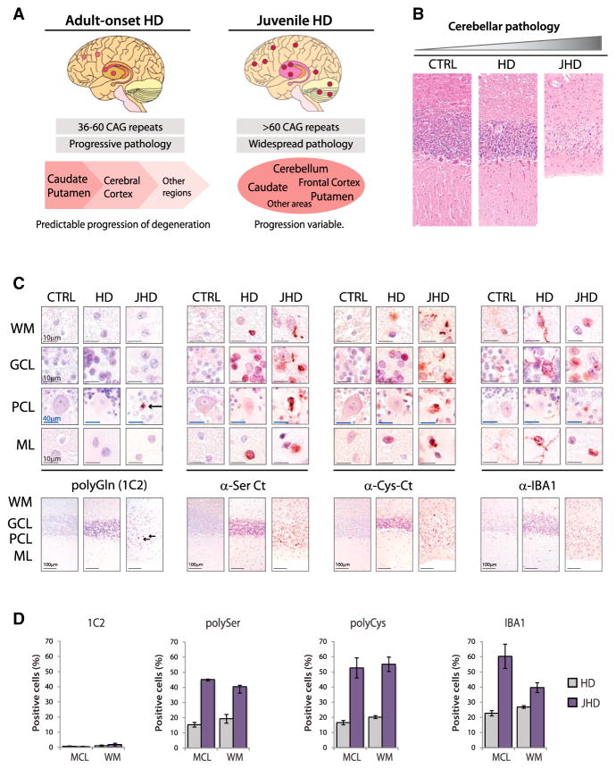
Figure 5. Increased RAN Protein Staining in Juvenile HD.
(A) Schematic diagram summarizing features of adult-onset and juvenile HD pathology.
(B) H&E staining in control, adult-onset, and juvenile-onset HD cases with cerebellar atrophy.
(C) α-polyGln, α-polySer, α-polyCys and α-IBA1staining in cerebellar layers.
(D) Quantitation of IHC-positive cells with 1C2 (polyGln) and α-polySer-Ct, α-polyCys-Ct, and α-IBA1 antibodies. WM, white matter; GCL, granule cell layer; PCL, Purkinje-cell layer; ML, molecular layer. Red, positive staining; blue, nuclear counterstain. Staining of cerebellum in adult-onset HD cases is variable. IHC images and quantification of percent positive cells represent typical positive regions. See also Figures S5A and S5B.

