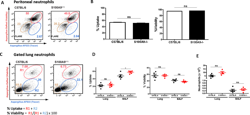
Figure 5. Conidia killing by neutrophils is independent of calprotectin.
A. Representative dot plots of peritoneal C57BL/6 or S100A9−/− neutrophils incubated with FLARE (dsRed+ AF633+) conidia 8h. B. Uptake and viability of FLARE conidia incubated with C57BL/6 or S100A9−/− neutrophils in vitro for 8 hrs. Representative In vitro experiments with 3–6 technical replicates per condition, significance was determined by student’s t-test. C57BL/6 or S100A9−/− bone marrow chimeras were challenged intratracheally with 3 × 10⁷ FLARE conidia (C-E). C. Representative dot plots of neutrophils from lung and BALF analyzed for dsRed and AF633 fluorescence by flow cytometry. The tan gates indicate bystander neutrophils, and the red (R1) and blue (R2) gates indicate neutrophils that contain live or killed conidia, respectively. D. Uptake and viability of FLARE conidia in lung or BALF neutrophils 12h pi. E. Total neutrophils in lung or BALF 12h pi in C57BL/6 or S100A9−/− bone marrow chimeras. In vivo experiments were repeated twice with at least 3 mice per group. Significance was determined by Mann-Whitney U test. All data show mean +/− SEM. See also Figure S3.

