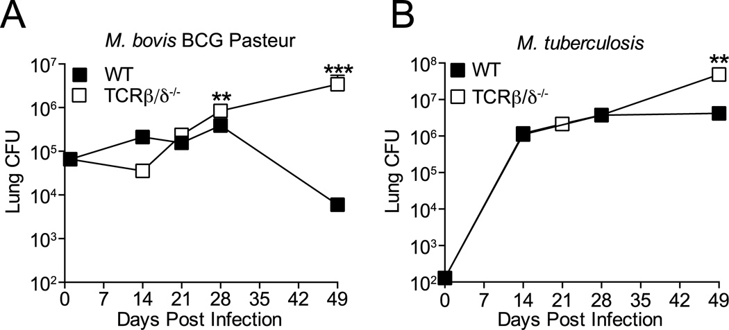
Figure 1.
T cells clear M. bovis BCG but not M. tuberculosis infection in the lungs. T cell-deficient (TCRβ/δ−/−) or WT C57BL/6 mice were infected with 5 × 10⁴ BCG or 100 M. tuberculosis H37Rv CFU by aerosol, and live bacteria were quantitated in the lungs for 7 weeks post infection. (A) M. bovis BCG. (B) M. tuberculosis H37Rv. Data are expressed as mean ± SEM, n = 5 mice per time point. Student t test comparing bacterial CFU in WT and TCRβ/δ−/− mice at each time point; *** P=0.0002, ** P =0.0078 The data shown are representative of 2 similar experiments.

