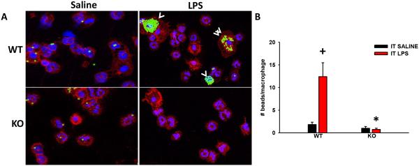
Figure 6. TRPV4 mediates LPS-stimulated macrophage phagocytosis of IgG-coated latex beads in vivo.
WT and TRPV4 KO C57BL/6 were treated with IT LPS (3 µg/g) for 16 h followed by intratracheal (IT) IgG-coated latex beads for 6 h. Cell phagocytic analysis was performed on the BAL by microscopic analysis of cytospin preparations. (A) Representative confocal images of WT and TRPV4 KO mice given IT saline (n = 2) or LPS (n = 5) followed by IgG-coated latex beads (white arrow heads) - Green-beads, Blue-nuclei (PMN: multilobular nucleus, macrophage: single concentric nucleus), Red-CD45 to show membrane (All panels, 40X Orig. Mag.). (B) LPS treated WT mice had increased macrophage phagocytosis of IgG-coated latex beads compared to the LPS treated TRPV4 KO. The number of beads per macrophage were quantified from panel A (*,+p < 0.05). + denotes the increase in LPS vs UT, * denotes difference in LPS response between KO and WT.

