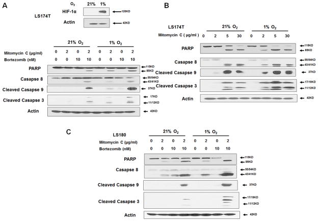
Figure 2. The combination of mitomycin C and bortezomib induced significant activation of caspases under hypoxia.
(A) LS174T cells were exposed to normoxia and hypoxia for 24 h and the intracellular level of hypoxia-inducible factor-1α (HIF-1α) was detected with immunoblotting (inset). LS174T cells were treated with mitomycin C (2 μg/ml) and/or bortezomib (10 nM) under normoxia and hypoxia for 24 h and caspase 8, cleaved caspase 9, and cleaved caspase 3 were detected with immunoblotting (main). (B) LS174T cells were treated with mitomycin C (2, 5, 30 μg/ml) under normoxia and hypoxia for 24 h and caspase 8, cleaved caspase 9, and cleaved caspase 3 were detected with immunoblotting. (C) LS180 cells were treated with mitomycin C (2 μg/ml) and bortezomib (10 nM) under normoxia and hypoxia for 24 h and caspase 8, cleaved caspase 9, and cleaved caspase 3 were detected with immunoblotting. Actin was used to confirm equal amounts of proteins loaded in each lane.

