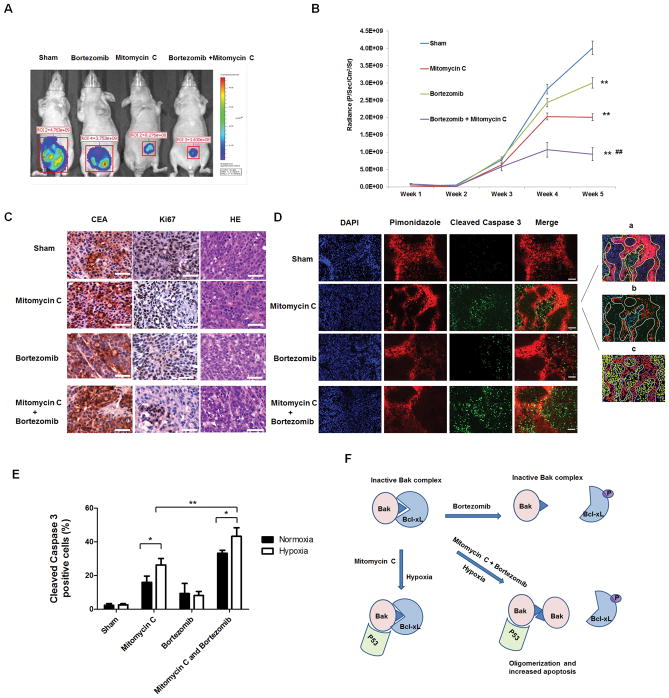
Figure 6. Effect of mitomycin C and bortezomib on the growth of LS174T intraperitoneal tumors.
(A) Nude mice were intraperitoneally (i.p.) inoculated with 5×10⁵ LS174T-luc cells/mouse on day 0. Four days after tumor inoculation, all the tumor-bearing mice were treated with either phosphate buffered saline (PBS) alone (sham) or bortezomib alone at a dose of (0.25 mg/kg body weight) or mitomycin C (1.5 mg/kg body weight) alone or the combination of mitomycin C and bortezomib every other day from day 4 to day 28. Mice were imaged using the IVIS Imaging System Series 200. ** represents a statistically significant difference (P<0.01) compared with the control group. ## represents a statistically significant difference (P<0.01) compared with the single treatment group (B) Line graph illustrating the luciferase activity (photons/second) in LS174T tumor-bearing mice treated with PBS alone, bortezomib alone, mitomycin C alone, or the combination from week 1 to week 5. (C) Tumor tissues were harvested at week 5 and subjected to H&E staining and immunohistochemistry staining with anti-CEA and ki67 primary antibodies. Representative images are shown (magnification, X400). Scale bar 50 μm. (D) Animals were sacrificed 1 h after pimonidazole injection (i.p.). Tissue was stained for cleaved caspase 3 (green) and pimonidazole adducts (red). Cell nuclei were stained with DAPI. Representative images are shown (magnification, X200). Scale bar 100 μm. Quantification was performed with Photoshop software: (a) Hypoxic region was determined by the yellow shape. (b) Cleaved caspase 3-positive cells were quantifed. (c) Cell nuclei were quantified. (E) Cleaved caspase 3-positive cells in normoxic and hypoxic areas were quantified. *P<0.05; ** P<0.01. (G) A schematic diagram of the mechanism of the combinatorial treatment with mitomycin C and bortezomib-induced apoptosis under hypoxia.

