
Figure 2. Distribution patterns of key neuronal cell populations in the amygdala.
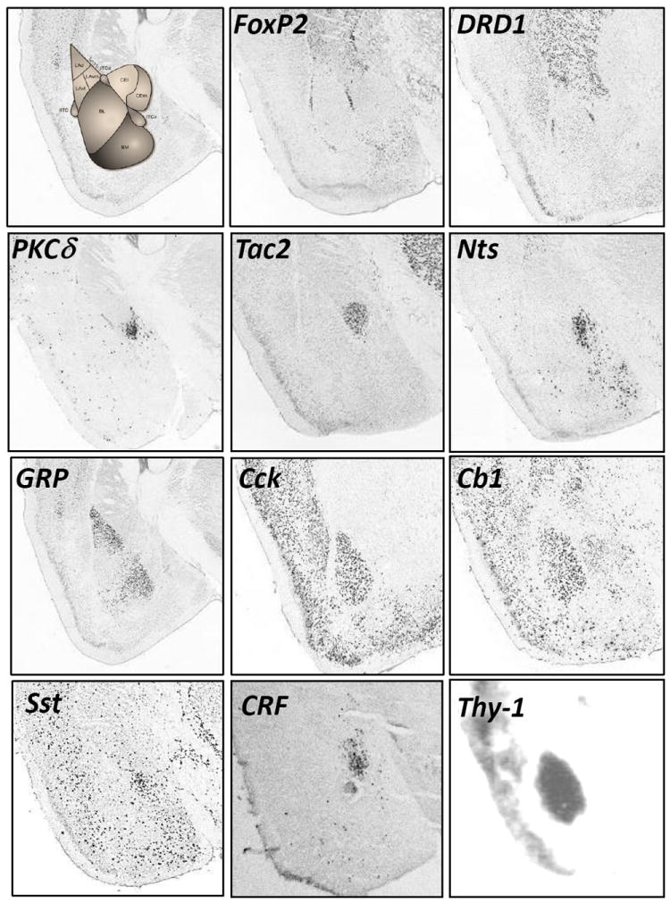
Distribution patterns of FoxP2 (Forkhead box protein P2), DRD1 (Dopamine receptor D1), PKCδ (Protein kinase C delta), Tac2 (Tachykinin 2), Nts (Neurotensin), GRP (Calcitonin Gene Related Peptide), CCK (Cholecystokinin), Cb1 (Cannabinoid receptor type 1), Sst (Somatostatin), CRF (Corticotropin Releasing Factor), Thy1 (Thy1 cell surface antigen) in the amygdala. Images from in situ hybridizations from Allen Brain Atlas as well as Ressler lab.