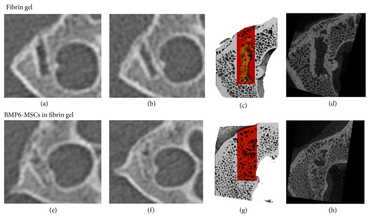
Figure 3.
BMP6-MSCs induce vertebral bone repair. Bone regeneration in lumbar vertebral defects was monitored using clinical CT imaging on 6 ((a) and (e)) and 12 weeks after surgery ((b) and (f)). Animals were sacrificed on week 24 (i.e., 6 months) after surgery and excised vertebrae were subjected to μCT imaging. Bone formation was quantified based on μCT scans (analyzed region is highlighted in red, (c) and (g)). Marked differences in bone regeneration can be seen in defects treated with BMP6-MSCs ((g) and (h)) versus fibrin gel only ((c) and (d)).

