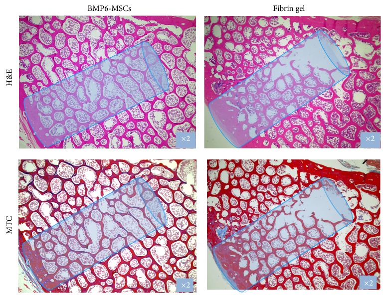
Figure 5.
Histological sections of vertebral bone defects. Treated vertebrae were harvested and processed for histology. Sections were stained with H&E and Masson's Trichrome and imaged using a light microscope; representative images showing advanced defect closure in the BMP6-MSC group versus the control (fibrin gel only) group.

