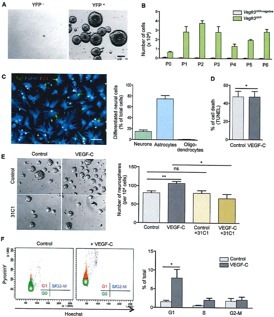
Figure 2. VEGF-C/VEGFR3 Signaling Activates Hippocampal NSCs In Vitro.
(A and B) Neurosphere cultures derived from sorted Vegfr3YFP and Vegfr3YFP-negativecells. The formation of neurospheres was only observed in Vegfr3YFP cell cultures that can self-renew for at least six successive passages.
(C) Representative images and quantification of neurosphere differentiation. Vegfr3YFP cells differentiate into TuJ1+ neuron (green), GFAP+ astrocyte (blue), and very few 04+ oligodendrocyte (red).
(D) Cell death was quantified by TUNEL staining.
(E) Representative images and quantification of neurospheres after treatment with VEGF-C (50 ng/ml) and a VEGFR3-function-blocking antibody (31C1).
(F) FACS profile and cell cycle analysis of control Vegfr3YFP cells and VEGF-C-treated Vegfr3YFP cells after Pyronin Y/Hoechst 33342 staining. The scale bars represent 50 µm (A and C).
Student’s t test: p < 0.05 (*); p < 0.005 (**); not significant (ns). Bars: mean ± SEM; n = 3–5 independent experiments. See also Figure S2.

