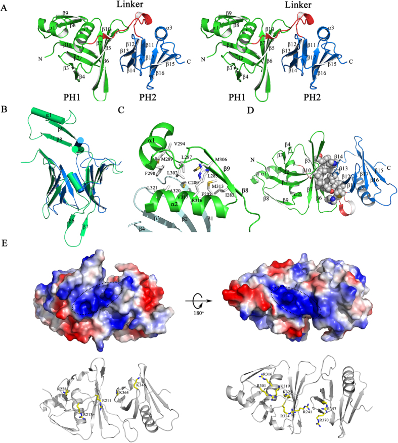
Figure 2. Structural features of SSRP1-M.
(A) A stereoscopic representation of SSRP1-M. The secondary structural elements are labelled. The structure is coloured according to the domains, as follows: the PH1 domain (residues 197–323), green; the inter-domain linker (residues 324–340), red; and the PH2 domain (residues 341–427), blue. (B) The structural superposition of PH1 domain (green) and PH2 domain (blue) of SSRP1-M with the differences labelled. (C) The extra βαβ topology of the PH1 domain stabilized by hydrophobic interactions. (D) The hydrophobic contacts between the PH1 domain and PH2 domain. The involved residues are indicated as spheres coloured by atom-type (O, red; N, blue; C and H, grey). (E) Upper panel, the electronic potential surface of SSRP1-M. The two positively charged patches are highlighted. Lower panel, the basic residues comprising the positively charged patches.

