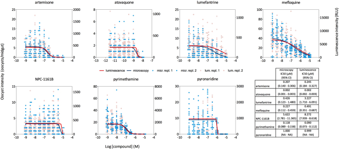
Figure 2. Standard Membrane Feeding Experiments for 7 compounds from the MMV validation box, assessed both by microscopy and luciferase assay.
All compounds were tested in nine dilutions in duplicate. The figure shows oocyst counts in individual mosquitoes and relative light units (RLU) from a distinct cohort of mosquitoes from the same cage. The table shows IC50 values and cognate 95% confidence intervals (CI) determined from fitting the data to a logistic regression model by Maximum Likelihood Estimation. NA: confidence interval not computed as the model did not converge.

