
Figure 7. LPS from pathogenic and commensal bacteria evade detection by CD14 and TLR4.
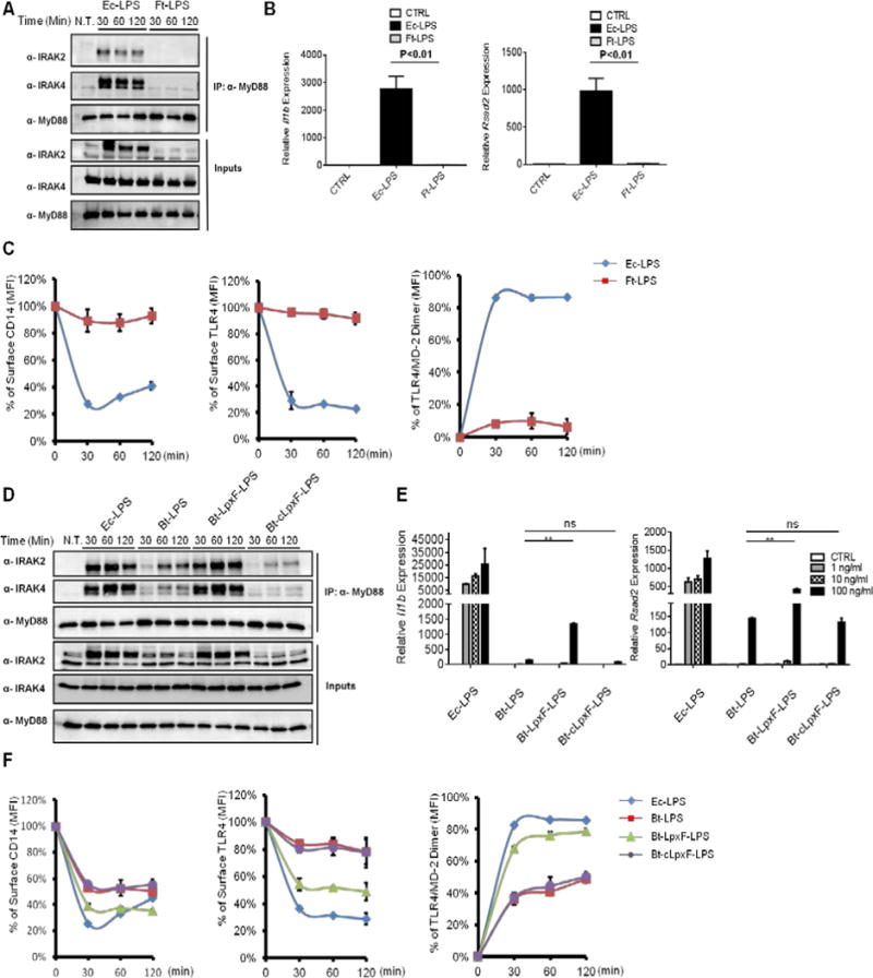
(A) iBMDMs were treated with Ec-LPS or Ft-LPS (1 μg/ml) for the indicated times, and the assembly of myddosome was examined as described in Figure 3B.
(B) iBMDMs were treated with Ec-LPS (100 ng/ml) or Ft-LPS (100 ng/ml) for 4 hours, and the expression of Il-1 beta (left) and rsad2 (right) was measured by qPCR.
(C–D) WT iBMDMs were stimulated with Ec-LPS or Ft-LPS (1 μg/ml) for the times indicated. CD14 endocytosis (left), TLR4 endocytosis (middle) and TLR4/MD-2 dimerization (right) were determined by flow cytometry (C) or myddosome was examined (D).
(E) Cells were treated with Ec-LPS and Bt-LPS species at the concentration of 1 ng/ml, 10ng/ml and 100 ng/ml for 4 hours, il1b and rsad2 expression was measured by qPCR.
(F) iBMDMs were stimulated with the LPS variants indicated (1 μg/ml). At the indicated times, CD14 endocytosis (left), TLR4 endocytosis (middle) and TLR4/MD-2 dimerization (right) were determined by flow cytometry.
Error bars represent mean SEM from triplicate readings in one experiment. **,p<0.01.