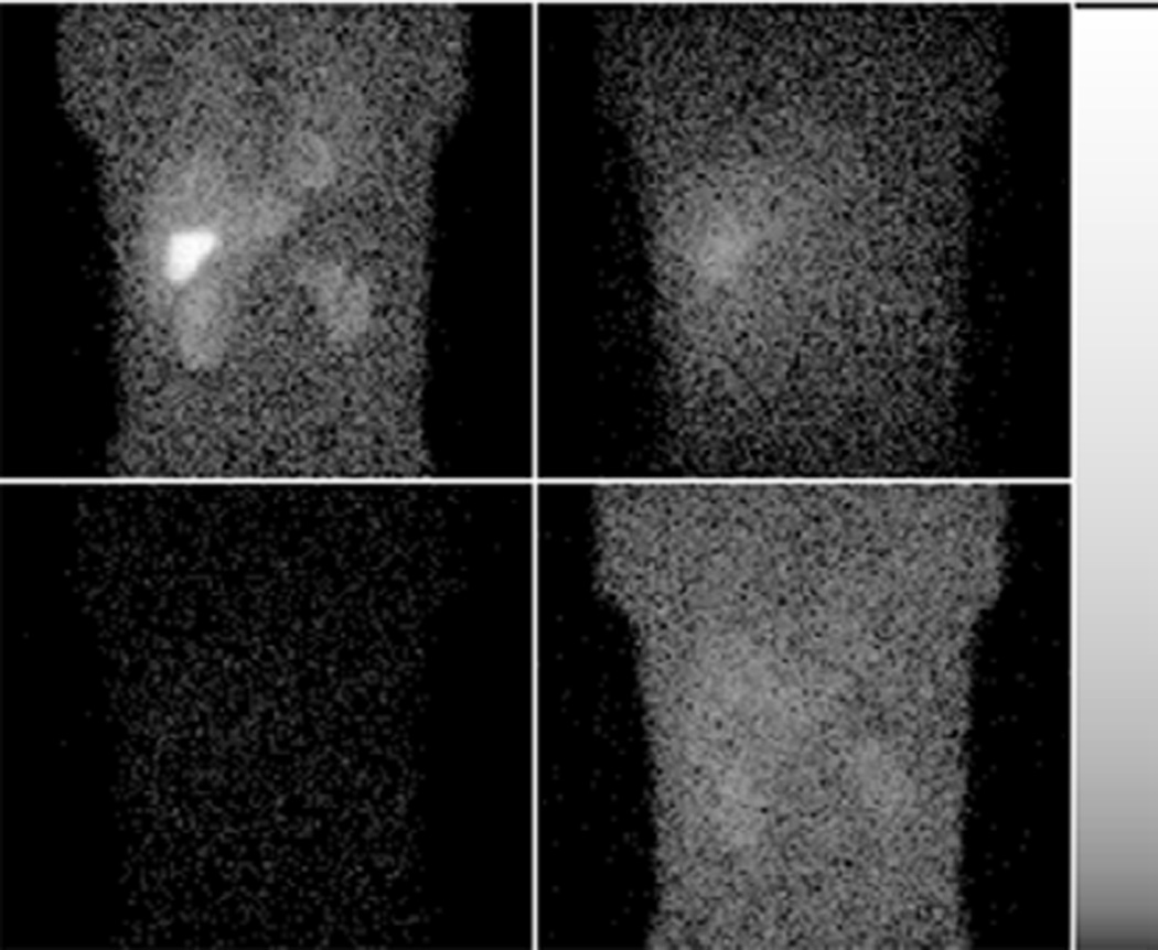
Figure 4.
Sample noisy projection images for Tc-99m (top row) and Tl-201 (bottom row) acquired in a 20% wide Tc-99m energy window (left) and 28% wide Tl-201 energy window, W5 (right). Activities of Tc-99m and Tl-201 correspond to configuration R3. Images are displayed on a logarithmic gray scale to show more clearly the lower uptake organs.

