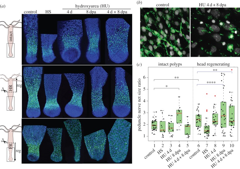
Figure 4.
Loss of basal neurogenesis in Hydra after HU exposure. (a) Anatomy of the basal nervous system detected with anti-RFamide immunostaining (green) in intact polyps (upper), in lower halves having regenerated their apical region for 8 days (middle) or in upper halves having regenerated their basal region for 8 days (lower). Conditions of HU treatment are shown in figure 2d. (b) Higher magnification of the basal nerve net in untreated and HU-treated (4 d + 8 dpa) intact animals. (c) Modulations of the RFamide basal index in intact and head-regenerating polyps. Black brackets indicate the statistical testing on mean values calculated with two-sided Welch t-test and blue brackets indicate statistical testing on the variance between two populations calculated with the F-test (*p < 0.05; **p < 0.01; ****p < 0.0001).

