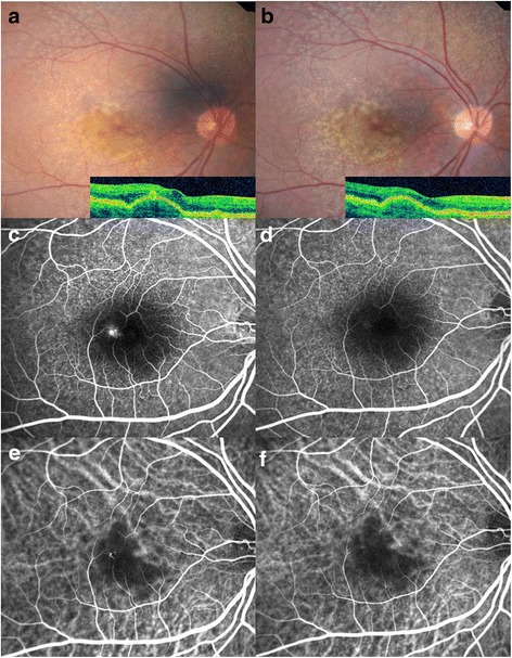
Fig. 5.

a Baseline fundus photograph and corresponding OCT image of a 62-year-old woman with retinal angiomatous proliferation (RAP) before injections. It demonstrates intraretinal neovascularization (IRN) with multiple drusen in the region of the fovea. The initial BCVA was 0.398 (LogMAR). b 12 months after injection, IRN decreased. The final BCVA was 0.201 (LogMAR). c FA shows a focal area of hyperfluorescence (leakage) corresponding to the IRN. d After injections, leakage in FA resolved in the IRN. e ICGA shows a focal area of intense hyperfluorescence or a so-called ‘hot spot’ corresponding to the lesion. f One year after treatment, ICGA showed resolution of hyperfluorescence
