Figure 1.
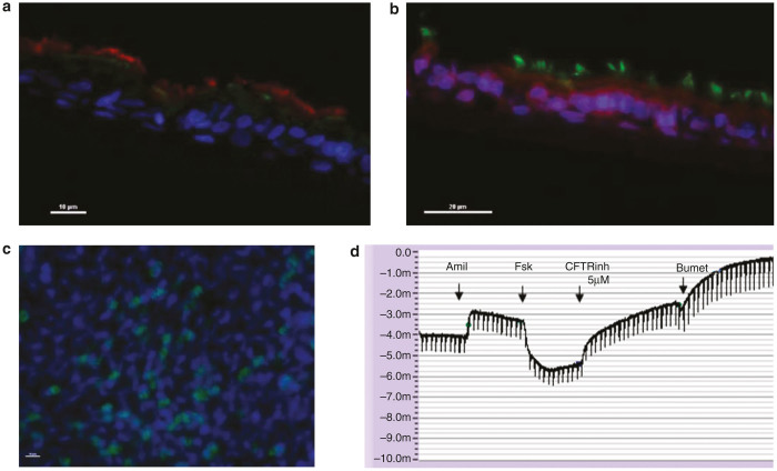
Primary culture of nasal epithelial cells results in a well-differentiated phenotype. Nasal cells were obtained by nasal brushing from non-cystic fibrosis (CF) human donor as described in methods. Epithelial cells were expanded in submerged cultures and then grown in air-liquid interface for 3 weeks. Cells imaged with confocal microscope. (a) Cross sectional view of human nasal epithelial cells demonstrating apical CFTR. Blue stain, DAPI (4’, 6-diamidino-2-phenylindole); red stain, β4 tubulin (cilia); green stain, CFTR protein. Bar indicates 10 µm. (b) Cross sectional view of human nasal epithelial cells demonstrating tight junctions. Blue stain, DAPI (looks purple); green stain, β4 tubulin (cilia); red stain, ZO1 (tight junctions). Bar indicates 20 µm. (c) Apical view of human nasal epithelial cells demonstrating cilia. Blue stain, DAPI; green stain, β4 tubulin (cilia). Bar indicates 10 µm. (d) Original Ussing trace from non-CF nasal cells displaying adequate bioelectric properties (Vte = −3.17 mV, Rte= 597 Ω cm2, Ieq = −5.31 µA/cm2) as well as functional expression of ENaC- and CFTR-mediated ion transport. CFTR, CF transmembrane conductance regulator; ENaC, epithelial sodium channel.

