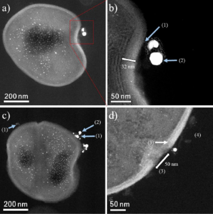
Figure 7.
HAADF-STEM micrographs of MRSA cells. (a) MRSA cells surrounded by AgNPs, with AgNPs smaller than 10 nm also being found inside of the cells. (b,c) (1) CWGs. (2) AgNPs interacting with WTAs and CWGs. MRSA cell micrograph shows a cell wall size of 32 nm. The Ag nanoparticle concentration is 23 ppm. (d) (3) Membrane destabilization (≈50 nm).

