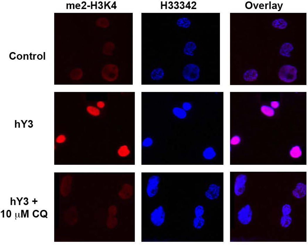
Figure 1. Differential in vitro expression of H3K4me2 antigen in THP-1 macrophages.
Resting and hY3 (a proxy of anti-Ro60 immune complex) stimulated macrophages with and without chloroquine (10 uM) were probed with an α-H3K4me2 antibody (red) and nuclei counterstain (blue). Note that from merged images, magenta indicates an overlap of H3K4me2 and the nucleus.

